Track progress, take quizzes and save notes on this lesson.

Free forever · no card needed

Start free
Intermediate

Microscopy and Magnification

4.1.1.2 Animal and plant cells (Required practical 1)·4.1.1.5 Microscopy·4.1.1.1 Eukaryotes and prokaryotes (orders of magnitude and standard form)

Aligned to the AQA 8461 specification

Level
Intermediate
Reading time
9 min
Published
16 June 2026
Updated
1 July 2026
On this page
  1. 1.Why Cells Need a Microscope
  2. 2.The Magnification Equation
  3. 3.Rearranging to Find Actual or Image Size
  4. 4.Units, Prefixes and Conversions
  5. 5.Standard Form for Very Small Sizes
  6. 6.Light vs Electron Microscopes
  7. 7.Required Practical 1: Observing Cells
  8. 8.Common Exam Mistakes

Key takeaways

  • Magnification is how many times bigger the image is than the real object, while resolution is the smallest distance at which two points can still be seen as separate.
  • Magnification = image size ÷ actual size; it has no units and the image and actual sizes must be in the same unit before dividing.
  • The key unit chain is 1 mm = 1000 µm = 1,000,000 nm; going to a smaller unit you multiply and to a larger unit you divide.
  • In standard form a number is written between 1 and 10 times a power of ten, and small lengths take a negative power, for example 0.0007 mm is 7 x 10⁻⁴ mm.
  • An electron microscope has higher magnification and higher resolution than a light microscope, which is why it reveals sub-cellular structures a light microscope cannot.

Why Cells Need a Microscope

Most cells are far too small to see with the naked eye. A typical animal cell is about 0.02 mm across — roughly the width of a fine hair split into many strands — so studying cells means magnifying them.

Two measurements describe how good a microscope is, and they are not the same thing:

  • Magnification — how many times bigger the image is than the real object.
  • Resolution (resolving power) — the smallest distance between two points that can still be seen as two separate points, rather than blurring into one.

A microscope that magnifies hugely but has poor resolution just gives you a bigger blur. To see fine detail you need both: high magnification and high resolution.

Resolution is the ability to distinguish two close objects as separate. It sets the real limit on how much useful detail you can see.

This is the idea the whole topic rests on, so be clear on the difference before moving to the equation.

The Magnification Equation

The relationship between magnification, the image size you measure, and the real size of the object is a single equation you must be able to use in both directions.

Magnification has no units — it is a ratio of two lengths, so it is written as a number with a "×", such as 400×. The image size and the actual size must be in the same unit before you divide.

Worked example — find the magnification

An image of a cell measures 50 mm across. The real cell is 0.05 mm across. Find the magnification.

StepWorking
Write the equationmagnification = image size ÷ actual size
Substitute (same units)magnification = 50 mm ÷ 0.05 mm
Calculatemagnification = 1000×

Check: 0.05 mm × 1000 = 50 mm, which matches the image. The answer is correct.

Rearranging to Find Actual or Image Size

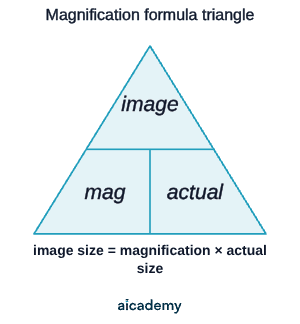
The same equation rearranges two ways. A formula triangle makes the rearrangement reliable: cover the quantity you want, and what is left tells you whether to multiply or divide.
Worked example — find the actual size

A cell is viewed at a magnification of 400×. Its image measures 24 mm across. Find the real size of the cell.

StepWorking
Rearrangeactual size = image size ÷ magnification
Substituteactual size = 24 mm ÷ 400
Calculateactual size = 0.06 mm
Convert to µm0.06 mm × 1000 = 60 µm

Check: 60 µm = 0.06 mm, and 0.06 mm × 400 = 24 mm, which matches the image. The answer is correct.

Decide what the question is asking for before you start: the magnification (a number), or a real length (which needs a sensible unit such as µm or nm).

Units, Prefixes and Conversions

Cells are measured in millimetres (mm), micrometres (µm) and nanometres (nm). Getting the prefixes and conversions right is where most marks are lost, so learn this ladder.

PrefixSymbolMetresRelationship
centicm m1 cm = 10 mm
millimm m1 mm = 1000 µm
microµm m1 µm = 1000 nm
nanonm m1 nm = m

The key chain to memorise:

Worked example — convert a small length

An organelle measures 0.0007 mm. Express this in micrometres and in nanometres.

StepWorking
mm → µm (× 1000)0.0007 mm × 1000 = 0.7 µm
µm → nm (× 1000)0.7 µm × 1000 = 700 nm

To go from a larger unit to a smaller one you multiply (the number gets bigger); going the other way you divide. Here 0.0007 mm and 700 nm are the same length written two ways.

Studying this for an exam?

Generate a personalised learning path for this subject. Free to get started.

Create a learning path

Standard Form for Very Small Sizes

Cell sizes often come out as awkward decimals like 0.0007 mm. Writing them in standard form — a number between 1 and 10 multiplied by a power of 10 — keeps them tidy and avoids miscounting zeros. The 8461 specification asks you to express answers in standard form where appropriate.

For a small number the power of 10 is negative; the power tells you how many places the decimal point moves.

Worked example — write a size in standard form

Express 0.0007 mm in standard form.

StepWorking
Move the decimal to get 1 ≤ a < 100.0007 → 7.0
Count the places moved (right)4 places
Negative power for a small number mm

So 0.0007 mm = mm. A bacterium about 0.002 mm long is mm, and a 60 µm cell is m. Orders of magnitude compare sizes by powers of ten: an object that is longer than another is one order of magnitude bigger, and a cell at m is about three orders of magnitude larger than an organelle at m.

(Higher Tier note — Foundation candidates handle standard form too, but more demanding standard-form manipulation and order-of-magnitude comparisons are most likely to appear on Higher papers.)

Light vs Electron Microscopes

A light microscope focuses visible light through glass lenses. An electron microscope fires a beam of electrons instead of light. Because electrons have a far shorter wavelength than light, an electron microscope resolves much finer detail — so it has both much higher magnification and much higher resolving power. That higher resolution is the reason it can reveal sub-cellular structures a light microscope cannot.

FeatureLight microscopeElectron microscope
Magnificationup to about 1500–2000×up to about 1,000,000× or more
Resolutionabout 200 nmabout 0.2 nm
Detail visiblenucleus, cell wall, chloroplasts, larger structuresinternal detail of organelles and sub-cellular structures
Specimensliving or deaddead only (samples are in a vacuum)
Cost / sizecheap, portable, used in schoolsvery expensive, large, specialist labs

The development of the electron microscope let biologists see and understand many more sub-cellular structures, because of its higher magnification and resolving power. That single point — better resolution reveals more detail — is the spec's headline for this topic.

The trade-off: a light microscope can show living cells, but only an electron microscope reveals the finest internal detail.

Required Practical 1: Observing Cells

Required practical 1 uses a light microscope to observe, draw and label plant and animal cells. You need to know the method and the drawing conventions, and a magnification scale must be included.

Slide preparation (e.g. onion epidermis or cheek cells):

  • Peel a thin, single-cell-thick layer of tissue and place it on a clean glass slide.
  • Add a drop of stain (such as iodine for plant cells or methylene blue for cheek cells) to make structures show up clearly.
  • Lower a coverslip at an angle with a mounting needle to avoid trapping air bubbles.

Focusing — start on low power:

  • Begin with the lowest-power objective lens, then move up to higher powers once the cells are in view.
  • Focus using the coarse adjustment first, then the fine adjustment for a sharp image.

Drawing conventions:

  • Use a sharp pencil with clear, continuous lines — no shading or colouring in.
  • Draw what you actually see, not a textbook diagram.
  • Use straight label lines that touch the structure, with labels written horizontally.
  • State the magnification used, and include a magnification scale.

Total magnification = eyepiece magnification × objective magnification. A ×10 eyepiece with a ×40 objective gives 10 × 40 = 400×.

Common Exam Mistakes

1. Confusing magnification with resolution

These are different. Magnification is how many times bigger the image is; resolution is the smallest detail you can still tell apart. Electron microscopes beat light microscopes on both, and it is the higher resolution that lets us see more sub-cellular detail.

2. Not converting to the same unit before dividing

The magnification equation only works if image size and actual size share a unit. Convert first (for example 0.05 mm stays in mm, or change everything to µm), then divide.

3. Forgetting the formula triangle direction

Magnification = image ÷ actual. To find the real size, divide the image by the magnification; to find the image size, multiply the magnification by the actual size. Cover the quantity you want in the triangle to check.

4. Getting the power of ten the wrong way round

Small lengths (less than 1) use a negative power in standard form, such as mm. Large numbers use a positive power. Count the decimal places carefully — a single miscounted zero loses the mark.

5. Shading or colouring biological drawings

In Required practical 1 drawings, use clean pencil lines only, with straight label lines and the magnification stated. Shading and colouring lose marks and are not part of the scientific drawing convention.

Key terms

Magnification
How many times bigger the image is than the real object, calculated as image size divided by actual size.
Resolution
The smallest distance between two points that can still be seen as two separate points rather than blurring into one.
Light microscope
A microscope that focuses visible light through glass lenses and can show living or dead specimens.
Electron microscope
A microscope that fires a beam of electrons, giving much higher magnification and resolution but only able to view dead specimens in a vacuum.
Standard form
A way of writing numbers as a value between 1 and 10 multiplied by a power of ten.
Order of magnitude
A comparison of sizes by powers of ten, where an object ten times longer than another is one order of magnitude bigger.
Micrometre (µm)
A unit of length equal to one thousandth of a millimetre, where 1 mm = 1000 µm.
Nanometre (nm)
A unit of length where 1 µm = 1000 nm, so 1 mm = 1,000,000 nm.

Frequently asked questions

Magnification is how many times bigger the image is than the real object, whereas resolution is the smallest distance between two points that can still be seen as separate. Electron microscopes beat light microscopes on both, and it is the higher resolution that reveals more sub-cellular detail.

Use magnification = image size ÷ actual size. Magnification has no units and is written as a number with a ×, such as 400×. The image size and actual size must be converted to the same unit before you divide.

An electron microscope fires a beam of electrons, which have a far shorter wavelength than light, so it has much higher resolving power as well as higher magnification. That higher resolution lets it reveal sub-cellular structures a light microscope cannot show.

Generate revision on any topic you study

Type any topic you're studying and Aicademy generates a complete lesson, quiz, and flashcard set, personalised to your level.

Lessons on anything

Structured, level-matched lessons on any topic you study

Practice quizzes

Find out what you actually know before the exam does

Flashcard sets

Lock in key concepts with instant revision cards

Ask Aica

Stuck on something? Get a clear explanation, any time

Prev

Cell Structure: Eukaryotes and Prokaryotes

Next

Mitosis and the Cell Cycle

Related lessons

7 min

Lesson

Culturing Microorganisms

AQA GCSE Biology · AQA 8461

1 month ago

Top students don’t revise more. They revise what counts.

Start revising free

Free to start. No card needed.