Track progress, take quizzes and save notes on this lesson.

Free forever · no card needed

Start free
Intermediate

The Heart and Blood Vessels

4.2.2.2 The heart and blood vessels

Aligned to the AQA 8461 specification

Level
Intermediate
Reading time
9 min
Published
16 June 2026
Updated
1 July 2026
On this page
  1. 1.How the Heart Is Built
  2. 2.The Vessels Attached to the Heart
  3. 3.Tracing Blood Through the Double Circuit
  4. 4.Why a Double Circuit Is Better
  5. 5.How the Heartbeat Is Controlled
  6. 6.Arteries, Veins and Capillaries
  7. 7.Identifying a Vessel From Its Features
  8. 8.Common Exam Mistakes

Key takeaways

  • The heart has four chambers: two atria on top that receive blood and two ventricles below that pump it out into the large vessels.
  • In a double circulatory system blood passes through the heart twice per circuit, once on the pulmonary loop to the lungs and once on the systemic loop to the body.
  • The left ventricle has a thicker muscular wall than the right because it pumps blood at high pressure all around the body, while the right only pumps the short distance to the lungs.
  • The pulmonary artery carries deoxygenated blood and the pulmonary vein carries oxygenated blood, the opposite of the usual artery/vein oxygen pattern.
  • Resting heart rate is set by a group of cells in the right atrium acting as a natural pacemaker; an artificial pacemaker is an electrical device fitted to correct an irregular heartbeat.

How the Heart Is Built

The heart is an organ made mostly of muscle. Its job is to pump blood around the body. It has four chambers: two at the top called atria (singular: atrium) and two below called ventricles.

The atria receive blood arriving at the heart. The ventricles pump blood out. Blood always flows in one direction — atria fill first, then push blood down into the ventricles, which contract to force it out into the large vessels.
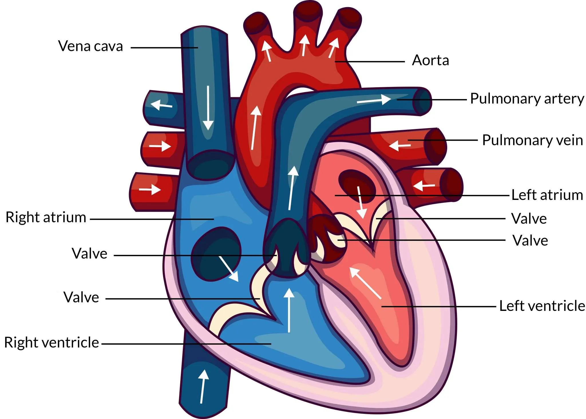
Cross-section of the human heart with the four chambers, valves and major blood vessels labelled

The heart is a double pump: the right side and left side work side by side at the same time, but push blood to two different destinations.

The Vessels Attached to the Heart

Four named blood vessels connect the heart to the rest of the body, plus the coronary arteries that feed the heart muscle itself. AQA only requires these names — you do not need to name the valves.

VesselCarries bloodConnected toOxygen level
Vena cavaInto the heart, from the bodyRight atriumDeoxygenated
Pulmonary arteryOut of the heart, to the lungsRight ventricleDeoxygenated
Pulmonary veinInto the heart, from the lungsLeft atriumOxygenated
AortaOut of the heart, to the bodyLeft ventricleOxygenated

The pulmonary vessels are the exception to the usual rule: the pulmonary artery carries deoxygenated blood and the pulmonary vein carries oxygenated blood, because they connect to the lungs rather than the body.

The heart muscle needs its own oxygen supply. The coronary arteries branch off the aorta and run across the surface of the heart, delivering oxygenated blood to the muscle. If a coronary artery becomes blocked, that section of muscle is starved of oxygen and can be damaged.

Valves sit between the atria and ventricles, and at the exits to the arteries. They snap shut to stop blood flowing backwards, keeping it moving in one direction only.

Tracing Blood Through the Double Circuit

In a double circulatory system, blood passes through the heart twice for every full circuit of the body. There are two separate loops:

  • Pulmonary circuit — heart → lungs → heart (picks up oxygen)
  • Systemic circuit — heart → body → heart (delivers oxygen)

Worked example — follow one red blood cell. Start with a cell arriving from the body, low on oxygen:
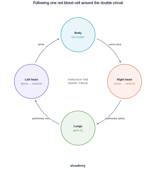
Step by step:

  1. Body (low oxygen)
  2. Vena cava
  3. Right atrium
  4. Right ventricle
  5. Pulmonary artery
  6. Lungs (gains oxygen, loses CO₂)
  7. Pulmonary vein
  8. Left atrium
  9. Left ventricle
  10. Aorta
  11. Body (delivers oxygen)

Steps 2–5 are the right side pumping deoxygenated blood to the lungs. Steps 7–10 are the left side pumping the now-oxygenated blood to the body. The cell went through the heart twice — once on each side.

Why a Double Circuit Is Better

A double circulatory system keeps oxygenated and deoxygenated blood completely separate, and lets the body return blood to the heart to be re-pressurised before the long journey to the tissues.

Blood loses pressure as it squeezes through the tiny vessels in the lungs. In a single circuit, that low-pressure blood would have to travel straight on to the rest of the body — slow and inefficient. The double circuit solves this:

FeatureBenefit
Blood returns to the heart after the lungsIt is pumped again at high pressure to the body
Oxygenated and deoxygenated blood kept apartBlood reaching tissues is fully oxygenated
Left ventricle pumps separately to the bodyPressure can be high enough to reach every cell

This is why the left ventricle has a much thicker muscular wall than the right: it must generate enough pressure to push blood all the way around the whole body, whereas the right ventricle only pumps the short distance to the nearby lungs.

Higher blood flow means tissues receive oxygen and glucose faster, supporting a high rate of respiration. This is essential for active, warm-blooded animals like humans.

Worth saving these ideas?

Turn what you've read into instant revision cards. Free to get started.

Make flashcards

How the Heartbeat Is Controlled

Heart muscle contracts on its own, but the resting heart rate is set by a group of cells in the right atrium that act as a natural pacemaker. These cells produce regular electrical impulses that spread across the heart and tell the muscle when to contract, keeping the beat steady and coordinated.

If this natural pacemaker stops working properly, the heartbeat can become too slow, too fast, or irregular. An artificial pacemaker can fix this:

An artificial pacemaker is a small electrical device, fitted under the skin, that sends regular electrical signals to the heart to correct an irregular heartbeat.

We can measure how hard the heart is working using cardiac output — the volume of blood pumped by a ventricle each minute:

Worked example. A person has a stroke volume (blood pushed out per beat) of 70 mL and a heart rate of 72 beats per minute:

During exercise the heart rate rises, so cardiac output rises and the muscles receive oxygen faster.

Arteries, Veins and Capillaries

The body has three types of blood vessel, each adapted to its job. The simple rule: arteries carry blood Away from the heart; veins carry blood back towards the heart.

FeatureArteryVeinCapillary
DirectionAway from heartTowards heartConnects arteries to veins
WallThick, muscular, elasticThinOne cell thick
Lumen (hole)NarrowWideVery narrow
Valves?NoYesNo
Blood pressureHighLowFalling
Main functionCarry blood at high pressureReturn blood at low pressureExchange substances with cells
  • Arteries have thick, elastic, muscular walls to withstand the high pressure of blood leaving the heart, and a narrow lumen to keep that pressure high.
  • Veins have thin walls and a wide lumen because the blood is at low pressure. They contain valves to stop blood flowing backwards on its slow journey back to the heart.
  • Capillaries are only one cell thick, so oxygen and glucose can diffuse out to cells and waste like CO₂ can diffuse in across a very short distance. Their walls are too thin to be felt as a pulse.

In the lungs, capillaries form a dense network around the alveoli (tiny air sacs), giving a huge, thin surface for gases to be exchanged between the air and the blood.

Identifying a Vessel From Its Features

Exams often describe a vessel and ask you to name it and justify your answer. Match the structure to the function.

Worked example — vessel A. "Has a thick muscular wall, a narrow lumen, no valves, and carries blood at high pressure."

  • Thick muscular wall + high pressure → it carries blood leaving the heart.
  • No valves and a narrow lumen fit this too.
  • Answer: an artery. Justification: thick muscular walls withstand the high pressure of blood being pumped away from the heart.

Worked example — vessel B. "Has a wall that is a single cell thick and connects small arteries to small veins."

  • A one-cell-thick wall is the giveaway.
  • It links arteries to veins and allows exchange.
  • Answer: a capillary. Justification: a wall one cell thick gives a short diffusion distance, so oxygen and glucose pass to cells and CO₂ passes into the blood.

A vessel with thin walls, a wide lumen and valves would be a vein — the valves and wide lumen suit low-pressure blood returning to the heart.

Justify your answer by linking structure to function. "It is an artery" earns little; "it is an artery because its thick muscular wall withstands high pressure" is much stronger.

Common Exam Mistakes

1. Saying the right ventricle has the thicker wall

It is the left ventricle that has the thicker, more muscular wall, because it pumps blood to the whole body. The right ventricle only pumps the short distance to the lungs, so its wall is thinner.

2. Mixing up the pulmonary vessels

The pulmonary artery carries deoxygenated blood (to the lungs) and the pulmonary vein carries oxygenated blood (from the lungs). This is the opposite of the usual artery/vein oxygen pattern — examiners test it often.

3. Confusing artery and vein direction

Arteries carry blood away from the heart (A for Away); veins carry blood back towards it. This is true regardless of whether the blood is oxygenated.

4. Forgetting why capillary walls are thin

A capillary wall is one cell thick so substances diffuse across a short distance. State the link to diffusion, not just "the wall is thin".

5. Describing the pacemaker as a muscle, not cells

The natural pacemaker is a group of cells in the right atrium that produce electrical impulses. An artificial pacemaker is a separate electrical device fitted to correct an irregular heart rate — do not confuse the two.

6. Saying blood passes through the heart once

In a double circulatory system blood passes through the heart twice per full circuit — once for the pulmonary (lung) loop and once for the systemic (body) loop.

Key terms

Atria
The two upper chambers of the heart that receive blood arriving at the heart.
Ventricles
The two lower chambers of the heart that contract to pump blood out into the large vessels.
Double circulatory system
A system in which blood passes through the heart twice for every full circuit of the body.
Pulmonary circuit
The loop carrying blood from the heart to the lungs and back to pick up oxygen.
Systemic circuit
The loop carrying blood from the heart to the body and back to deliver oxygen.
Coronary arteries
Arteries that branch off the aorta and deliver oxygenated blood to the heart muscle itself.
Natural pacemaker
A group of cells in the right atrium that produce electrical impulses to set the resting heart rate.
Artificial pacemaker
A small electrical device fitted under the skin that sends regular signals to correct an irregular heartbeat.
Cardiac output
The volume of blood pumped by a ventricle each minute, equal to stroke volume multiplied by heart rate.
Artery
A blood vessel with thick muscular elastic walls and a narrow lumen that carries blood away from the heart at high pressure.
Vein
A blood vessel with thin walls, a wide lumen and valves that returns blood towards the heart at low pressure.
Capillary
A blood vessel one cell thick that connects arteries to veins and allows substances to be exchanged with cells.

Frequently asked questions

The left ventricle has a thicker, more muscular wall because it must generate enough pressure to pump blood all the way around the whole body. The right ventricle only pumps the short distance to the nearby lungs, so its wall is thinner.

Arteries carry blood away from the heart and have thick muscular walls and a narrow lumen to withstand high pressure. Veins carry blood back towards the heart with thin walls, a wide lumen and valves. Capillaries are one cell thick for exchange with cells.

The natural pacemaker is a group of cells in the right atrium that produce regular electrical impulses to set the resting heart rate. An artificial pacemaker is a small electrical device fitted under the skin to correct an irregular heartbeat.

Generate revision on any topic you study

Type any topic you're studying and Aicademy generates a complete lesson, quiz, and flashcard set, personalised to your level.

Lessons on anything

Structured, level-matched lessons on any topic you study

Practice quizzes

Find out what you actually know before the exam does

Flashcard sets

Lock in key concepts with instant revision cards

Ask Aica

Stuck on something? Get a clear explanation, any time

Prev

The Digestive System and Food Tests

Next

Blood: Components and Functions

Related lessons

10 min

13 min

Lesson

Coronary Heart Disease

AQA GCSE Biology · AQA 8461

1 month ago

9 min

Top students don’t revise more. They revise what counts.

Start revising free

Free to start. No card needed.