Track progress, take quizzes and save notes on this lesson.

Free forever · no card needed

Start free
Intermediate

The Eye

4.5.2.3 The eye (Biology only)

Aligned to the AQA 8461 specification

Level
Intermediate
Reading time
12 min
Published
16 June 2026
Updated
1 July 2026
On this page
  1. 1.The Eye as a Sense Organ
  2. 2.Structures of the Eye
  3. 3.The Pupil Reflex
  4. 4.Accommodation: Focusing the Lens
  5. 5.Defects of the Eye: Myopia and Hyperopia
  6. 6.Worked Example: Choosing the Correcting Lens
  7. 7.New Technologies for Correcting Vision
  8. 8.Common Exam Mistakes

Key takeaways

  • The eye is a sense organ whose receptors in the retina are sensitive to light intensity and colour and send impulses along the optic nerve to the brain.
  • Accommodation is changing the shape of the lens to focus on near or distant objects; for a near object the ciliary muscles contract, suspensory ligaments loosen and the lens becomes thicker and more rounded.
  • The pupil reflex is automatic: in bright light the pupil constricts to protect the retina, and in dim light it dilates to let more light in, controlled by the iris.
  • In myopia (short sightedness) light focuses in front of the retina and is corrected with a concave lens; in hyperopia (long sightedness) light focuses behind the retina and is corrected with a convex lens.
  • Vision can also be corrected by contact lenses, laser surgery that reshapes the cornea, or replacing the eye's natural lens with an artificial one.

The Eye as a Sense Organ

The eye is a sense organ: it contains receptors that detect a stimulus and convert it into electrical impulses for the nervous system. The receptors in the eye are sensitive to light intensity (how bright the light is) and colour.

These light receptors sit in the retina at the back of the eye. When light lands on them they send impulses along the optic nerve to the brain, which interprets them as an image.

To see clearly, the eye must do two jobs:

  • Focus light from objects at different distances onto the retina — this is called accommodation.
  • Control how much light enters so the retina is neither overwhelmed in bright light nor starved in dim light — this is the job of the iris and pupil.

A sense organ is a group of receptor cells that respond to a specific type of stimulus. The eye responds to light.

The rest of this lesson works through the named structures, how the eye focuses, how it adjusts to changing light, and the two common focusing defects you are expected to know.

Structures of the Eye

You must be able to identify these structures on a diagram and explain how each structure is related to its function. Below is a labelled cross-section through the eye, viewed from the side, with light entering from the left.
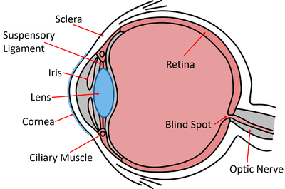
Cross-section of the human eye with the cornea, pupil, iris, lens, ciliary muscle, sclera, retina and optic nerve labelled

Light passes in this order: cornea → pupil (the gap in the iris) → lens → retina.

StructureFunction (how its structure suits its job)
ScleraTough, white outer layer; protects the eye and keeps its shape.
CorneaTransparent front layer; refracts (bends) light rays — it does most of the focusing as light enters.
IrisColoured ring of muscle; controls the size of the pupil and so the amount of light entering.
PupilThe hole in the centre of the iris that light passes through (not a structure itself — it is the gap).
LensTransparent, flexible disc; changes shape to fine-tune focus onto the retina (accommodation).
Ciliary musclesRing of muscle that contracts or relaxes to change the lens shape.
Suspensory ligamentsConnect the ciliary muscles to the lens; transmit the pull that flattens the lens.
RetinaLight-sensitive layer at the back; contains receptors sensitive to light intensity and colour.
Optic nerveCarries electrical impulses from the retina to the brain.

The Pupil Reflex

The iris adjusts the size of the pupil to control how much light reaches the retina. This is a reflex — automatic and rapid — which protects the receptors in bright light and lets the eye gather as much light as possible in dim light.

The iris contains two sets of muscle that work as antagonistic pairs (one set contracts while the other relaxes):

ConditionsWhat happensResult
Bright lightCircular muscles contract, radial muscles relaxPupil gets smaller — less light enters, protecting the retina
Dim lightRadial muscles contract, circular muscles relaxPupil gets larger — more light enters so the eye can still detect an image

In bright light the pupil constricts (gets smaller); in dim light it dilates (gets larger). The change is automatic — you do not decide to do it.

The pupil reflex is the eye's way of adapting to light intensity. Adapting to dim light keeps enough light reaching the receptors so a faint image can still be formed.

(Extra context — the two muscle types are the circular and radial muscles of the iris. AQA 8461 requires you to know the iris controls pupil size as a reflex, but does not name these two muscle sets in the specification.)

Accommodation: Focusing the Lens

Accommodation is the process of changing the shape of the lens to focus light from near or distant objects onto the retina. The ciliary muscles and suspensory ligaments work together to do this.

The key idea: the ciliary muscles and suspensory ligaments have an opposite (inverse) relationship with the lens shape.

Ciliary musclesSuspensory ligamentsLens shapeRefraction
Near objectContractLoosen (slacken)Thicker / more roundedRefracts light strongly
Distant objectRelaxPulled tightThinner / flatterRefracts light only slightly

The logic of the inverse relationship:

  • When the ciliary muscles contract, the ring of muscle gets smaller, so it stops pulling on the suspensory ligaments. The ligaments slacken, the lens is released, and it springs into a fatter, more curved shape. A fatter lens bends light more — needed for near objects.
  • When the ciliary muscles relax, the ring widens, pulling the suspensory ligaments tight. The taut ligaments stretch the lens thin and flat. A flatter lens bends light less — needed for distant objects, whose rays arrive almost parallel and need little bending.

Remember the trap: ciliary muscles contracting makes the lens fatter, not thinner. Contraction releases the lens rather than squeezing it.

Worked walk-through — focusing on a near object. A student lifts a phone close to their face to read a message. Step through what each part does, in order:

Step 1 — identify the distance. The object is near, so the lens must become a strong refractor to bend the light enough to land on the retina.

Step 2 — the ciliary muscles. They contract, making the ring of ciliary muscle smaller.

Step 3 — the suspensory ligaments. Because the muscle ring is now smaller, the suspensory ligaments loosen (go slack). They stop pulling on the edge of the lens.

Step 4 — the lens. Released from the pull, the elastic lens becomes thicker and more curved (more rounded).

Step 5 — the result. The thicker lens refracts the light strongly, bringing the rays from the close object to a focus exactly on the retina — a sharp image forms.

Reverse every step for the same student looking up at a distant tree: ciliary muscles relax → suspensory ligaments pulled tight → lens stretched thin and flat → light refracted only slightly → distant rays focus on the retina.

Exam shortcut: name the four stages in order — ciliary muscles → suspensory ligaments → lens shape → refraction — and get the direction right for each.

How much of this have you taken in?

Quiz yourself on this section, free, no card needed.

Test myself

Defects of the Eye: Myopia and Hyperopia

In a healthy eye, light focuses exactly on the retina. In the two common defects, the rays do not focus on the retina, so the image is blurred. These are usually corrected with spectacle lenses that refract the light so it focuses back on the retina.

The two defects are mirror images of each other:

DefectCommon nameProblem with focusCauseCorrecting spectacle lens
MyopiaShort sightednessLight focuses in front of the retinaEyeball too long, or the lens stays too curved — distant objects look blurredConcave (diverging) lens
HyperopiaLong sightednessLight focuses behind the retinaEyeball too short, or the lens too flat / inflexible — near objects look blurredConvex (converging) lens

How each correcting lens works:

  • A concave lens spreads the rays apart (diverges them) before they enter the eye. The eye then focuses them slightly further back — onto the retina instead of in front of it. This corrects myopia.
  • A convex lens brings the rays together (converges them) before they enter the eye. The eye then focuses them slightly further forward — onto the retina instead of behind it. This corrects hyperopia.

Memory hook: myopia → concave (both have an "o"-ish gap; the lens is thin in the middle and pushes the focus point back). Hyperopia → convex (thick in the middle, pulls the focus point forward).

Worked Example: Choosing the Correcting Lens

Question: A person can read a book held close to them clearly, but road signs in the distance look blurred. Identify the defect, state where the light is focusing, and choose the spectacle lens that corrects it. Justify your choice.

Step 1 — match the symptom to the defect. Distant objects are blurred while near objects are clear → this is myopia (short sightedness).

Step 2 — state where the light focuses. In myopia the rays from a distant object converge in front of the retina, so the image is out of focus by the time it reaches the receptors.

Step 3 — choose the lens. A concave (diverging) lens is needed.

Step 4 — justify it. The concave lens spreads the light rays apart before they reach the cornea and lens. This pushes the focus point further back, onto the retina, so the distant object appears sharp.

Now the opposite case — "near print is blurred but distant objects are clear":

  • Symptom → near blurred → hyperopia (long sightedness).
  • Light focuses behind the retina.
  • Correct with a convex (converging) lens, which brings the rays together first so the focus point moves forward onto the retina.

Quick check before you commit: blurred distance = myopia = concave; blurred near = hyperopia = convex.

New Technologies for Correcting Vision

Spectacle lenses are the traditional correction, but the spec also expects you to know newer methods. Each works by changing how the light is refracted so it focuses on the retina.

TechnologyHow it works
Contact lensesThin lenses (hard or soft) that sit directly on the surface of the cornea, refracting light like spectacles but less visible and convenient for sport.
Laser surgeryA laser changes the shape of the cornea, altering how strongly it refracts light so the image focuses on the retina without lenses.
Replacement lensThe eye's natural lens is removed and an artificial lens is implanted inside the eye to correct the focusing.

Each option trades off cost, convenience and risk. For example, contact lenses avoid the inconvenience of glasses but carry a small risk of eye infection, while laser surgery and replacement-lens surgery are permanent but involve the usual risks of an operation.

For the exam, link each technology back to the same goal: refracting the light so it focuses on the retina. Laser surgery is the one that changes the cornea; the replacement lens swaps out the eye's own lens.

Common Exam Mistakes

1. Getting the ciliary muscle direction backwards

For a near object the ciliary muscles contract and the lens becomes fatter / more rounded. Many students wrongly say the muscles contract to flatten the lens. Contraction releases the lens so it rounds up; relaxation tightens the ligaments and flattens it.

2. Mixing up myopia and hyperopia corrections

Myopia (short sight) → concave lens; hyperopia (long sight) → convex lens. Tie it to where the light focuses: in front of the retina (myopia) needs the rays spreading out (concave); behind the retina (hyperopia) needs them brought together (convex).

3. Calling the pupil a structure

The pupil is the gap in the centre of the iris, not a piece of tissue. It is the iris (a muscle) that changes the pupil's size.

4. Confusing the cornea and the lens

The cornea does most of the refraction as light first enters and its shape is fixed. The lens fine-tunes the focus by changing shape during accommodation. Laser surgery reshapes the cornea; a replacement-lens operation swaps the lens.

5. Saying the iris reflex is a conscious choice

The pupil reflex is automatic. In bright light the pupil constricts to protect the retina; in dim light it dilates to let more light in. You do not control it deliberately.

6. Forgetting which way the suspensory ligaments move

The ligaments and ciliary muscles have an inverse relationship. Ciliary muscles contract → ligaments loosen. Ciliary muscles relax → ligaments are pulled tight. State both the muscle action and the ligament action together — describing only one half often leaves a description answer incomplete.

Key terms

Sense organ
A group of receptor cells that respond to a specific type of stimulus; the eye responds to light.
Retina
The light-sensitive layer at the back of the eye containing receptors sensitive to light intensity and colour.
Cornea
The transparent front layer of the eye that refracts (bends) light rays and does most of the focusing as light enters.
Iris
A coloured ring of muscle that controls the size of the pupil and so the amount of light entering the eye.
Pupil
The hole in the centre of the iris that light passes through.
Lens
A transparent, flexible disc that changes shape to fine-tune focus onto the retina during accommodation.
Ciliary muscles
A ring of muscle that contracts or relaxes to change the shape of the lens.
Suspensory ligaments
Ligaments connecting the ciliary muscles to the lens that transmit the pull which flattens it.
Accommodation
The process of changing the shape of the lens to focus light from near or distant objects onto the retina.
Myopia
Short sightedness, where light focuses in front of the retina so distant objects look blurred; corrected with a concave lens.
Hyperopia
Long sightedness, where light focuses behind the retina so near objects look blurred; corrected with a convex lens.
Optic nerve
The nerve that carries electrical impulses from the retina to the brain.

Frequently asked questions

For a near object the ciliary muscles contract, the suspensory ligaments loosen, and the lens becomes thicker and more rounded. This more curved lens refracts the light strongly so it focuses exactly on the retina. The reverse happens for distant objects.

Myopia (short sightedness) is corrected with a concave (diverging) lens that spreads the rays apart so they focus on the retina instead of in front of it. Hyperopia (long sightedness) is corrected with a convex (converging) lens that brings the rays together.

No. The pupil is the hole in the centre of the iris that light passes through, not a piece of tissue. It is the iris, a coloured ring of muscle, that changes the pupil's size to control how much light enters the eye.

Generate revision on any topic you study

Type any topic you're studying and Aicademy generates a complete lesson, quiz, and flashcard set, personalised to your level.

Lessons on anything

Structured, level-matched lessons on any topic you study

Practice quizzes

Find out what you actually know before the exam does

Flashcard sets

Lock in key concepts with instant revision cards

Ask Aica

Stuck on something? Get a clear explanation, any time

Next

Control of Body Temperature

Related lessons

10 min

10 min

Lesson

The Brain

AQA GCSE Biology · AQA 8461

1 month ago

Top students don’t revise more. They revise what counts.

Start revising free

Free to start. No card needed.