Track progress, take quizzes and save notes on this lesson.

Free forever · no card needed

Start free
Intermediate

The Brain

4.5.2.2 The brain (Biology only)

Aligned to the AQA 8461 specification

Level
Intermediate
Reading time
10 min
Published
16 June 2026
Updated
1 July 2026
On this page
  1. 1.What the Brain Does and What It Is Made Of
  2. 2.The Cerebral Cortex
  3. 3.The Cerebellum and the Medulla
  4. 4.How Neuroscientists Study the Brain
  5. 5.Why Studying Brain Damage Maps Function
  6. 6.Why the Brain Is So Difficult to Investigate and Treat
  7. 7.Common Exam Mistakes

Key takeaways

  • The brain controls complex behaviour, is made of billions of interconnected neurones, and is organised into different regions that each carry out a different function.
  • The cerebral cortex is the folded outer layer responsible for consciousness, intelligence, memory and language.
  • The cerebellum coordinates muscular activity (movement) and balance, while the medulla controls unconscious activities such as heart rate and breathing.
  • Neuroscientists map brain regions to functions by studying patients with brain damage, electrically stimulating parts of the brain, and using MRI scanning.
  • Investigating and treating the brain is difficult because it is complex (billions of interconnected neurones) and delicate, so procedures on it are risky.

What the Brain Does and What It Is Made Of

The brain controls complex behaviour. It interprets information from receptors, makes decisions, and sends instructions to effectors — but it also handles thought, memory and language, which go far beyond a simple reflex.

The brain is made of billions of interconnected neurones. A neurone is a nerve cell that carries electrical impulses. Because these neurones are wired together in vast numbers, the brain can process huge amounts of information at once.

The brain is not uniform. It has different regions that carry out different functions. For GCSE you must be able to identify three regions on a diagram of the brain and describe what each one does: the cerebral cortex, the cerebellum and the medulla.
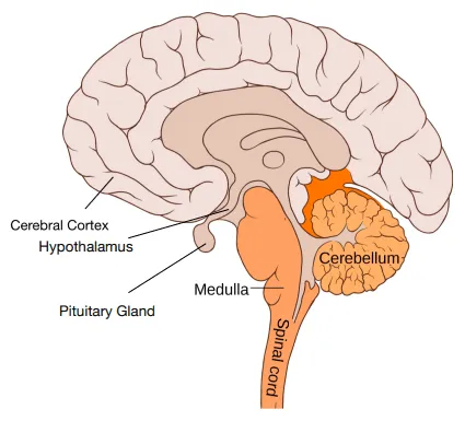
Side view of the human brain showing the cerebral cortex, cerebellum and medulla labelled

Key idea: the brain controls complex behaviour. It is made of billions of interconnected neurones, organised into different regions, each carrying out a different function.

The Cerebral Cortex

The cerebral cortex is the large, highly folded outer layer of the brain. It is the region responsible for our most complex, conscious abilities.

The cerebral cortex is responsible for:

  • Consciousness — being awake and aware of your surroundings.
  • Intelligence — reasoning and solving problems.
  • Memory — storing and recalling information.
  • Language — understanding and producing speech and writing.

A useful way to remember these four functions is the phrase "C-I-M-L": Consciousness, Intelligence, Memory, Language.

Key definition — cerebral cortex: the outer layer of the brain responsible for consciousness, intelligence, memory and language.

Applied walk-through — a patient who cannot form new memories after surgery:

  1. The deficit is the loss of memory — the patient cannot store new information.
  2. Memory is one of the four functions of the cerebral cortex.
  3. So the damage is most likely to the cerebral cortex, not the cerebellum (movement) or medulla (heart rate and breathing).
  4. Justification: the cerebellum coordinates muscular activity and balance, and the medulla controls unconscious activities — neither stores memories — so by elimination, and because memory is a cortical function, the cerebral cortex is the affected region.

The Cerebellum and the Medulla

Two more regions complete the three you must know. They control movement and the body's automatic processes respectively.

The cerebellum sits below and behind the cerebral cortex. It is responsible for the coordination of muscular activity (movement) and balance. It does not decide to move — it makes movements smooth, accurate and balanced.

The medulla is at the base of the brain, where it joins the spinal cord. It controls unconscious activities — things your body does without you thinking about them, such as heart rate and breathing.

RegionLocationMain function
Cerebral cortexLarge folded outer layerConsciousness, intelligence, memory, language
CerebellumLower back of the brainCoordination of muscular activity (movement) and balance
MedullaBase of the brain, joining the spinal cordControlling unconscious activities such as heart rate and breathing

Be precise: the cerebellum coordinates movement and balance — it does not control breathing. Breathing and heart rate are the medulla's job. Mixing these two up is the most common error on this topic.

Applied walk-through — a person with damage who becomes clumsy and unsteady on their feet:

  1. The symptoms are poor coordination of movement and loss of balance.
  2. These are exactly the functions of the cerebellum.
  3. So the damaged region is the cerebellum. The cerebral cortex would affect memory/language; the medulla would affect heart rate or breathing — neither matches "clumsy and unsteady".

How Neuroscientists Study the Brain

Neuroscientists have mapped regions of the brain to particular functions using three main methods. You should be able to describe all three.

(Higher Tier only — the Higher Tier specifically requires you to explain how these methods are used to map function and why brain investigation is difficult.)

  1. Studying patients with brain damage. When a small, specific region is damaged — by injury, a stroke or a tumour — doctors record which abilities the patient loses. If damage to one region reliably removes one ability, that region must be involved in that function.
  2. Electrically stimulating different parts of the brain. A small electrical current applied to a region triggers a response — a movement, a sensation, or a memory. The response tells researchers what that part of the brain does.
  3. Using MRI scanning techniques. MRI (magnetic resonance imaging) produces detailed images of the brain. Scanning while a person performs a task shows which regions become more active during that task.
MethodWhat it revealsLimitation
Studying brain damageFunction lost when a region stops workingRelies on accidental, often imprecise damage
Electrical stimulationResponse produced by stimulating a regionInvasive; usually only during brain surgery
MRI scanningWhich regions are active during a taskShows activity, not always cause

Exam tip: if asked "how have scientists mapped the brain?", name all three — studying patients with brain damage, electrical stimulation, and MRI scanning. One method on its own is an incomplete answer.

Something not quite clicking?

Ask Aica to explain any part of this differently. Free, takes 30 seconds.

Ask Aica

Why Studying Brain Damage Maps Function

Studying patients with brain damage is the oldest of the three methods, and the reasoning behind it is worth understanding rather than just memorising.

Applied walk-through — how a single case maps a function:

  1. A patient has a small, localised area of the cerebral cortex damaged in an accident.
  2. After the injury, the patient can no longer understand spoken language, but every other ability is intact.
  3. Because only that one region was damaged and only one ability was lost, neuroscientists infer that this region is responsible for understanding language.
  4. When many patients with damage to the same region all lose the same ability, the link between region and function becomes far more reliable.

This is why brain damage is so informative: it acts like a natural experiment — removing one part and revealing what stops working.

Key idea: by recording which functions are lost when a specific region is damaged, neuroscientists build a map linking each brain region to the ability it controls. Electrical stimulation and MRI then confirm and refine that map.

The reasoning has limits, though. Real injuries are rarely neat — damage often spreads across more than one region, so the lost ability cannot always be tied to a single spot. This is one reason the methods are used together rather than alone.

Why the Brain Is So Difficult to Investigate and Treat

(Higher Tier only — explaining the difficulties of investigating brain function and treating brain damage and disease is a Higher Tier requirement.)

The complexity and delicacy of the brain make investigating and treating brain disorders very difficult, and procedures on the brain are risky.

  • Complexity: the brain contains billions of interconnected neurones. A single function can involve many regions working together, so it is hard to pin one ability to one place.
  • Delicacy: brain tissue is soft, easily damaged, and does not heal like skin or bone. Even a small mistake during surgery can cause permanent harm.
  • Access: the brain is encased in the skull and protected by membranes, so reaching it for treatment means risky surgery.
  • Treating disease and damage: many drugs cannot cross from the blood into the brain (the blood–brain barrier blocks them), so delivering treatment is hard. Operating to remove a tumour risks damaging healthy regions nearby.

(Higher Tier only — you may be asked to evaluate the benefits and risks of a procedure on the brain or nervous system. A balanced answer weighs a benefit, e.g. removing a tumour could save a life, against a risk, e.g. nearby healthy tissue controlling speech or movement could be damaged, then gives a justified conclusion.)

Applied walk-through — evaluating surgery to remove a deep brain tumour (HT):

  1. Benefit: removing the tumour could relieve pressure and prolong or save the patient's life.
  2. Risk: the tumour sits near the medulla, which controls heart rate and breathing — damaging it during surgery could be fatal.
  3. Judgement: the decision depends on the tumour's exact position, the patient's condition, and whether safer alternatives (such as radiotherapy) exist. A good evaluation states a conclusion and justifies it, rather than just listing points.

(Extra context — not required by AQA 8461.) The blood–brain barrier is the named structure that stops many drugs reaching the brain. You can describe the effect — "many drugs cannot reach the brain" — without naming the barrier for 8461.

Common Exam Mistakes

1. Swapping the cerebellum and the medulla

The cerebellum coordinates movement and balance. The medulla controls unconscious activities such as heart rate and breathing. These are different regions with different jobs — do not give the cerebellum's job to the medulla or vice versa.

2. Saying the cerebral cortex "controls everything"

The cerebral cortex is responsible for consciousness, intelligence, memory and language — the conscious, higher functions. Movement coordination (cerebellum) and automatic processes like breathing (medulla) are not cortical functions.

3. Naming only one method for studying the brain

There are three: studying patients with brain damage, electrically stimulating parts of the brain, and MRI scanning. A question asking how the brain is studied expects more than one.

4. Confusing "controls" with "coordinates"

The cerebellum coordinates muscular activity — it makes movements smooth and balanced. It does not initiate or decide on movements. Use the right verb in your answer.

5. Treating "difficult to treat" as a vague statement

If asked why brain disorders are hard to investigate and treat, give specific reasons: the brain's complexity (billions of interconnected neurones, many regions involved in one function) and its delicacy (soft tissue that does not heal, hard to reach without risky surgery).

6. Giving a one-sided evaluation (HT)

When evaluating a brain procedure, you need both a benefit and a risk, followed by a justified conclusion. Listing only the benefits, or only the risks, does not meet what an "evaluate" question is asking for.

Key terms

Brain
The organ that controls complex behaviour; made of billions of interconnected neurones organised into regions with different functions.
Neurone
A nerve cell that carries electrical impulses.
Cerebral cortex
The large folded outer layer of the brain, responsible for consciousness, intelligence, memory and language.
Cerebellum
The region at the lower back of the brain responsible for coordinating muscular activity (movement) and balance.
Medulla
The region at the base of the brain, joining the spinal cord, that controls unconscious activities such as heart rate and breathing.
MRI scanning
Magnetic resonance imaging, which produces detailed images of the brain and shows which regions become more active during a task.

Frequently asked questions

The cerebral cortex, the cerebellum and the medulla. The cerebral cortex handles consciousness, intelligence, memory and language; the cerebellum coordinates movement and balance; the medulla controls unconscious activities such as heart rate and breathing.

Scientists map regions to functions using three main methods: studying patients with brain damage to see which abilities are lost, electrically stimulating different parts of the brain to trigger responses, and using MRI scanning to show which regions are active during a task.

The brain is extremely complex, with billions of interconnected neurones where one function can involve many regions, and it is delicate soft tissue that does not heal easily. It is encased in the skull, so reaching it means risky surgery.

Generate revision on any topic you study

Type any topic you're studying and Aicademy generates a complete lesson, quiz, and flashcard set, personalised to your level.

Lessons on anything

Structured, level-matched lessons on any topic you study

Practice quizzes

Find out what you actually know before the exam does

Flashcard sets

Lock in key concepts with instant revision cards

Ask Aica

Stuck on something? Get a clear explanation, any time

Prev

The Nervous System and Reflexes

Related lessons

10 min

12 min

Lesson

The Eye

AQA GCSE Biology · AQA 8461

1 month ago

Top students don’t revise more. They revise what counts.

Start revising free

Free to start. No card needed.